The distal end of the humerus, however, articulating with the radius and ulna in a fashion that no support is lent by any sort of contact with the body, is a ginglymus (hinge) joint and lateral motion, because of the long transverse diameter of its articular portions, is easily prevented by the medial and lateral ligaments (internal and external ligaments). Flexion of this, the humeroradioulnar joint (elbow), is restrained by the triceps brachii and extension is checked by the biceps brachii (flexor brachii).
The carpal joint (erroneously called the knee joint), is composed of the several carpal bones which interarticulate and, when taken as a group, serve as a means of attachment and articulation for the radius and metacarpal bones.
The transverse diameter of this joint is long, thus giving it contacting surfaces that are sufficiently extensive to minimize the strain upon the mesial and lateral ligaments (internal and external lateral common ligaments). Motion is that of flexion and extension; slight rotation is possible when the position is that of flexion. While supporting weight the carpus is fixed in position by a slight dorsal flexion, but undue dorsal flexion is prevented by the flexor muscles and tendons and volar-carpal or annular ligament, together with the superior check ligament.
The metacarpophalangeal articulation (fetlock joint), is a hinge joint and its articular surfaces contact one another, with respect to their having a long bearing surface from side to side, as do all ginglymus (hinge) joints. Two common lateral ligaments bind the bones together. While bearing weight, there is assumed a position of slight dorsal flexion, undue flexion being checked by the inhibitory apparatus of the joint—check ligaments, and their tendons and the suspensory ligament. The inhibitory apparatus of the fetlock joint is materially reinforced by the proximal sesamoid bones. Situated as they are, between the bifurcating portions of the suspensory ligament and the posterior part of the distal end of the metacarpus—with which they articulate—the sesamoid bones serve to change the course of the branches of the suspensory ligament in a manner that they give firm support to this joint. Volar flexion is limited by the extensors of the phalanges.
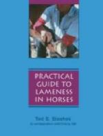
[Illustration: Fig. 4—Sagital Section of Digit and Distal Part of Metacarpus.
A, Metacarpal bone; B, first phalanx; C, second phalanx, D, third phalanx; E, distal sesamoid bone; 1, volar pouch of capsule of fetlock joint; 2, inter-sesamoidean ligament; 3, 4, proximal end of digital synovial sheath; 5, ring formed by superficial flexor tendon; 6, fibrous tissue underlying ergot; 7, ergot; 8, 9, 9’, branches of digital vessels; 10, distal ligament of distal sesamoid bone; 11, suspensory ligament of distal sesamoid bone; 12, 12’, proximal and distal ends of bursa podotrochlearis. (From Sisson’s “Anatomy of the Domestic Animals").]


