The femur may be considered analagous to the humerus in that it bears a similar relationship to the ilium, that exist between the humerus and scapula. Further flexion during repose is prevented chiefly by the glutens medius (maximus) muscle and its tendons. The larger tendon inserts to the summit of the trochanter major of the femur and corresponds to the biceps brachii in the action of the latter on the scapulohumeral joint, except that the gluteus medius, in attaching to the femoral trochanter, exerts its effect as a lever of the first class. Because of the relationship between the long axes of the femur and iliac shaft it is evident that the angle formed by these two bones is maintained chiefly by the gluteus muscles during weight bearing. Contraction of muscular fibers of the gluteus medius causes extension of the femur and muscular strain is prevented to a great degree by the inelastic portion of this muscle. The chief physiological antagonistics of the glutei are the quadriceps femoris and tensor fascia lata.
While the leg is supporting weight the stifle joint is fixed in position mainly by the quadriceps femoris group of muscles which are attached to the patella. Tendinous fibres intersect this muscular mass and relieve muscular strain during weight bearing. Because of the manner in which the patella functionates with the trochlea of the femur, comparatively little energy is required to prevent further flexion of the stifle joint. The patella, according to Strangeways, may be considered a sesamoid bone.
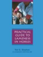
[Illustration: Fig. 45—Left stifle joint; front view. The capsules are removed. 1. Middle patellar ligament. 2. Stump of fascia lata. 3. Stump of common tendon of extensor longus and peroneus tertius. (From Sisson’s “Anatomy of Domestic Animals.")]
The quadriceps group of muscles is assisted by the anterior digital extensor (extensor pedis) peroneus tertius and tibialis anticus (flexor metatarsi) muscles. The latter pair (flexor metatarsi, muscular and tendinous portions, because of their attachment to the external condyle of the femur and to the metatarsal bone) are enabled to automatically flex the tarsal joint when the stifle is flexed.
The hock is kept fixed in position by the gastrocnemius and the superficial digital flexor (perforatus). The latter structure, which is chiefly tendinous, originates in the supracondyloid fossa of the femur and has an insertion to the summit of the fibular tarsal (calcis) bone. It relieves the gastrocnemius of muscular strain during weight bearing.
Smith[35] styles the function of the stifle and hock joints a reciprocating action, and we quote from this authority the following:


