Still, if we limit ourselves to a comparison of the same carbonized wood, preserved on the one hand by petrifaction, and on the other hand non-mineralized, we find a very perceptible diminution in bulk. The elements have contracted in length, breadth, and thickness, but principally in the direction of the compression that they have undergone in the purely carbonized specimens.
In the vicinity of the carbonized portions, those of the tracheae that have not done so have perceptibly preserved their primitive length, which has, so to speak, been maintained by their neighbors, but their other dimensions have become much smaller—a quarter in thickness and half in length.
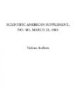
[Illustration: 12i: FIG. 9.—Calamodendron, Commentry; prosenchymatous portion of the wood carbonized, X200.]
If the two fragments of the same wood are, one of them silicified and the other simply carbonized and preserved in sandstone, the diminution in volume will have occurred in all directions in the latter of the two.
[Illustration: 12j: FIG. 10.—Calamodendron, fragment of the vascular portion of the wood carbonized.]
Figs. 9 and 11, which represent a portion of the fibrous region of Calamodendron wood, may give an idea of the shrinkage that has taken place therein. In Figs. 11 and 12, which show a few tracheae and medullary rays of the ligneous bands of the same plant, we observe the same phenomenon. We might cite a large number of analogous examples, but shall be content to give the following: Figs. 13 and 15 represent radial and tangential sections of the bark of Syringodendron pes-caprae. This is the first time that one has had before his eyes the anatomical structure of the bark of a Syringodendron, a plant which has not yet been found in a petrified state. It is coal, then, with its structure preserved, that allows of a verification of the theory advanced by several scientists that the often bulky trunks of Syringodendron are bases of Sigillariae.
[Illustration: 12k: FIG. 11.—Calamodendron, from Autun; prosenchymatous portion of the wood silicified, X200.]
[Illustration: 12l: FIG. 12.—Calamodendron, from Autun; vascular portion of the wood silicified.]
If we refer to Fig. 13, which represents a radial vertical section running through the center of one of the scars that permitted the specimen to be determined, we shall observe, in fact, a tissue formed of rectangular cells, longer than wide, arranged in horizontal series, and very analogous in their aspect to those that we have described in the suberose region of the bark of Sigillariae. Fig. 15 shows in tangential section the fibrous aspect of this tissue, which has been rendered denser through compression. Fig. 14 shows it restored. In Fig. 13, the external part of the bark is occupied by a thick layer of cellular tissue that exists over the entire surface of the


