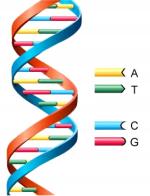
Deoxyribonucleic acid (DNA) is a double-stranded, helical molecule that forms the molecular basis for heredity.
For replication (duplication) to occur, DNA must first unwind, or "unzip," itself to allow the genetic information-encoding bases to become accessible. The base pairing within DNA is specific and complementary and, consequently, when the molecule unwinds, two complimentary strands are temporarily produced, each of which acts as a template for a new strand. At the onset of replication, a replication fork is created as the DNA molecule separates at a small region. The enzyme DNA polymerase then adds complementary nucleotides to each side of the freshly separated strands. The DNA polymerase adds nucleotides only to one end of the DNA. As a result, one strand (the leading strand) is replicated continuously, while the other strand (the lagging strand) is replicated discontinuously, in short bursts. Each of these small sections is finally joined to its neighbor by the action of another enzyme, DNA ligase, to yield a complete strand. The whole process gives rise to two completely new and identical daughter strands of DNA.
The discovery of the double-helix molecular structure of deoxyribonucleic acid (DNA) in 1953 was one of the major scientific events of the twentieth century, and marked the culmination of an intense search involving many scientists. Ultimately, credit for the discovery, and the 1962 Nobel Prize in the physiology or medicine category, went to James Dewey Watson, who at the time of the discovery was an American postdoctoral student from Indiana University; and Francis Harry Compton Crick, a researcher at the Cavendish Laboratory in Cambridge University, England. Their work, conducted at Cavendish Laboratory, significantly impacted the emerging field of molecular biology.
The double helix refers to DNA's spiral staircase structure, consisting of two right-handed helical polynucleotide chains coiled around a central axis. Genes, specific regions of DNA, contain the instructions for synthesizing every protein. Because life cannot exist without proteins, and the differences in proteins and protein enzymes control biological reactions, the discovery of DNA's structure unveiled one of the fundamental secrets of life: protein synthesis. In fact, the central dogma of molecular biology is that DNA is used to build ribonucleic acid, which is used to build proteins, which in turn play a role in building DNA and RNA.
Prior to Watson and Crick's discovery, it had long been known that DNA contained four kinds of nucleotides, which are the building blocks of nucleic acids, such as DNA and RNA. A nucleotide contains a five-carbon sugar called deoxyribose, a phosphate group, and one of four nitrogen-containing bases: adenine (A), guanine (G), thymine(T), and cytosine (C). Thymine and cytosine are smaller, single-ringed structures called pyrimidines; adenine and guanine are larger, double-ringed structures called purines. Watson and Crick drew upon this and other scientific knowledge in concluding that DNA's structure possessed two nucleotide strands twisted into a double helix, with bases arranged in pairs such as A T, T A, G C, C G. Along the entire length of DNA, the double-ringed adenine and guanine nucleotide bases were probably paired with the single-ringed thymine and cytosine bases. Using paper cutouts of the nucleotides, Watson and Crick shuffled and reshuffled combinations. Later, they used wires and metal to create their model of the twisting nucleotide strands that form the double-helix structure. According to Watson and Crick's model, the diameter of the double helix measures 2.0 nanometers (nm). Each turn of the helix is 3.4 nm long, with 10 bases in each chain making up a turn.
Before Watson and Crick's discovery, no one knew how hereditary material was duplicated prior to cell division. Using their model, it is now understood that enzymes can cause a region of a DNA molecule to unwind one nucleotide strand from the other, exposing bases that are then available to become paired up with free nucleotides stockpiled in cells. A half-old, half-new DNA strand is created in a process that is called semiconservative replication.
When free nucleotides pair up with exposed bases, they follow a base-pairing rule which requires that A always pairs with T, and G always with C. This rule is constant in DNA for all living things, but the order in which one base follows another in a nucleotide strand differs from species to species. Thus, Watson and Crick's double-helix model accounts for both the sameness and the immense variety of life.
Even though the long-term survival of species may be improved by genetic changes, the survival of individuals requires genetic stability. To achieve this, each living cell has a complement of enzymes whose function is to repair errors or damage in the DNA. Such lesions can arise spontaneously from normal cellular processes, (e.g., errors in replication) or can be generated by external factors such as chemicals or radiation. The altered portion of the DNA is recognized, usually because the DNA becomes distorted, and the damaged bases of sequences removed by nucleases. The correct sequence is then re-synthesized by a polymerase and the ends joined to the original DNA by a ligase. There are a number of enzyme systems that repair DNA damage and nearly all rely on the existence of two copies of the genetic information, one on each strand of the DNA double helix. Thus if the sequence on one strand is accidentally changed, the complementary strand still holds the correct information and can be used as a template to correct the alteration.
Watson and Crick's discovery of the double helix would not have been possible without significant prior discoveries. In his 1968 book, The Double Helix, A Personal Account of the Discovery of the Structure of DNA, Watson wrote that the race to unveil the mystery of DNA was chiefly a matter of five people: Maurice Wilkins, Rosalind Franklin, Linus Pauling, Crick, and Watson. Wilkins, an Irish biophysicist who shared the 1962 Nobel Prize in Physiology or Medicine with Crick and Watson, extracted DNA gel fibers and analyzed them, using x-ray diffraction. The diffraction showed a helical molecular structure, and Crick and Watson used that information in constructing their double-helix model. Franklin, working in Wilkins' laboratory, between 1950 and 1953, produced improved x-ray data using purified DNA samples, confirming that each helix turn is 3.4 nm. Although her work suggested DNA might have a helix structure, Franklin did not postulate a definite model. Pauling, an American chemist and twice Nobel laureate, in 1951 discovered the three-dimensional shape of the protein collagen. Pauling discovered that each collagen polypeptide or amino acid chain twists helically, and that the helical shape is held by hydrogen bonds. With Pauling's discovery, scientists worldwide began racing to discover the structure of other biological molecules, including the DNA molecule.
DNA fingerprinting, also called DNA profiling or genetic profiling, applies a test to determine the unique DNA sequence that each person carries for the purpose of identification. In the mid 1980s, Sir Alec Jeffreys at the University of Leicester coined the term DNA fingerprint and envisioned its powerful use. A single hair, a drop of blood, semen, or other body fluid can reveal the identity of a person. DNA fingerprinting is used for identifying people, studying populations, and forensic investigations.
Chromatin is a network of deoxyribonucleic acid (DNA) and nucleoproteins that constitutes a chromosome. Chromatin can only be found in a cell with a nucleus, and is therefore not present in a prokaryotic cell. The DNA within a eukaryotic cell can be as long as 12 cm (4.7 in). Due to its length, the DNA must be arranged and organized in order to fit within the small area of a cell nucleus. To accomplish this task, the DNA is bound, through electrostatic forces, with nucleoproteins called histones and nonhistones. The assemblage of DNA with the nucleoproteins is called a nucleosome, which is the fundamental structural unit of chromatin and represents 1.8 turns of DNA wound around a core particle of another histone protein. It is the nucleosomes, along with the DNA material between nucleosomes (linker segment), that gives DNA the characteristic beads-on-a string appearance, with the nucleosomes representing the bead and the linker segment of DNA representing the string.
When chromatin is isolated, it appears to be composed as smooth fibers. While the highest level of chromatin organization is not well understood, scientists have found that chromatin fibers are divided into functional groups, called domains. The domains are grouped and arranged into loops called solenoids. In cells that are dividing, the solenoids are further condensed into chromatids; an identical pair of chromatids comprise the recognizable shape of a chromosome.
There are two types of chromatin: heterochromatin and euchromatin. Heterochromatin is chromatin in condensed form, is seen as dense patches and is transcriptionally inactive while euchromatin is seen as delicate, thread-like structures that are abundant in active transcription cells.
This is the complete article, containing 1,446 words
(approx. 5 pages at 300 words per page).