Each pair of chromosomes in the eukaryotic cell contains a unique set of genes and thus the DNA sequence is also unique for each different chromosome pair. When chromosomes are condensed in metaphase, these differences in DNA content produce detectable differences in the staining pattern resulting from exposure to various dyes or stains that interact with the chromatin. The distinctive pattern of bands produced in this manner allows each chromosome pair to be identified and distinguished from all other chromosomes.
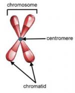
Prior to the use of banding methods, chromosomes could be distinguished only by comparing size and the position of the centromere. The first banding method for distinguishing human chromosomes was described by Swedish cytochemist T. O. Caspersson (1910-) in 1969 using the fluorescent dye, quinacrine. This method is called Q-banding. Regions of the genome which are relatively rich in the bases adenine and thymidine fluoresce brightly with Q-banding, and each human chromosome pair gives a unique pattern of bands. Since that time, Q-banding has been applied to a wide variety of other eukaryotic organisms.
Soon after the introduction of Q-banding, a wide variety of different stains were investigated for chromosome banding, including other fluorescent stains which bind to AT-rich regions (e.g. DAPI), and fluorescent stains which bind to GC-rich regions (eg. chromomycin-A). Because the pattern of banding is reversed with stains that bind to GC-rich regions, the method is called R-banding when these dyes are used.
The most frequently used method of banding for human cytogenetics involves trypsin digestion of the metaphase cells followed by staining with Giemsa, a dark blue stain which can be easily seen with standard light microscopy. This method is called G-banding. The banding pattern with G-banding is similar to that in Q-banding in that AT-rich regions of the genome stain more intensely than GC-rich regions. Alternatively, there are banding techniques available which produce an R-banding pattern using Giemsa stain but with different methods of pretreatment (e.g. high temperature incubation).
There are a variety of other banding methods that are used to characterize different aspects of chromatin structure and organization, or to identify subtle differences between the two homologs of a specific chromosome pair. These include methods for staining the nucleolar organizer regions or satellites of the acrocentric chromosomes, heterochromatin, or telomeres. These methods use various physical, chemical or enzymatic pretreatments to alter the structure of the chromatin including heat, ultraviolet light, barium hydroxide, protease treatment and restriction enzyme digestion.
This is the complete article, containing 403 words
(approx. 1 page at 300 words per page).