BookRags.com Literature Guides Literature
Guides
Criticism & Essays Criticism &
Essays
Questions & Answers Questions &
Answers
Lesson Plans Lesson
Plans
My Bibliography Periodic Table U.S. Presidents Shakespeare Sonnet Shake-Up
Research Anything:        
History | Encyclopedias | Films | News | Create a Bibliography | More... Login | Register | Help

Search "Chromosomal Mutations and Abnormalities"

Contents Navigation
 

Chromosomal Mutations and Abnormalities

Print-Friendly  Order the PDF version  Order the RTF version
About 7 pages (2,020 words)
Chromosome Summary

Bookmark and Share Questions on this topic? Just ask!

Chromosomal Mutations and Abnormalities

Chromosome abnormalities describe alterations in the normal number of chromosomes or structural problems within the chromosomes themselves. Both kinds of chromosome abnormalities may result from an egg or sperm with the incorrect number of chromosomes, or with a structurally faulty chromosome uniting with a normal egg or sperm during conception. Some chromosome abnormalities may occur shortly after conception. In this case, the zygote, the cell formed during conception that eventually develops into an embryo, divides incorrectly. Chromosomal abnormalities can cause serious mental or physical disabilities, and may lead to the death of the embryo. Zygotes that receive a full extra set of chromosomes, a condition called polyploidy, usually do not survive inside the uterus, and are spontaneously aborted (a process sometimes called a miscarriage).

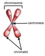
A chromosome consists of the body's genetic material, the deoxyribonucleic acid, or DNA, along with many kinds of protein. Within the chromosomes, the DNA is tightly coiled around these proteins (called histones) allowing huge DNA molecules to occupy a small space within the nucleus of the cell. When a cell is not dividing, the chromosomes are invisible within the cell's nucleus. Just prior to cell division, the chromosomes uncoil and begin to replicate. As they uncoil, the individual chromosomes look somewhat like a fuzzy X. Chromosomes contain the genes, or segments of DNA that encode for proteins, of an individual. When a chromosome is structurally faulty, or if a cell contains an abnormal number of chromosomes, the types and amounts of the proteins encoded by the genes is changed. When proteins are altered in the human body, the result can be serious mental and physical defects and disease.

Humans have 22 pairs of autosomal chromosomes and one pair of sex chromosomes, for a total of 46 chromosomes. These chromosomes can be studied by constructing a karyotype, or organized depiction, of the chromosomes. To construct a karyotype, a technician stops cell division just after the chromosomes have replicated using a chemical such as colchicine; the chromosomes are visible within the nucleus at this point. The chromosomes are photographed, and the technician cuts up the photograph and matches the chromosome pairs according to size, shape, and characteristic stripe patterns (called banding).

In most animals, two types of normal cell division exist. In mitosis, cells divide to produce two identical daughter cells. Each daughter cell has exactly the same number of chromosomes. This preservation of chromosome number is accomplished through the replication of the entire set of chromosomes just prior to mitosis.

Sex cells, such as eggs and sperm, undergo a different type of cell division called meiosis. Because sex cells each contribute half of a zygote's genetic material, sex cells must carry only half the full complement of chromosomes. This reduction in the number of chromosomes within sex cells is accomplished during two rounds of cell division, called meiosis I and meiosis II. Prior to meiosis I, the chromosomes replicate, and chromosome pairs are distributed to daughter cells. During meiosis II, however, these daughter cells divide without a prior replication of chromosomes. Mistakes can occur during either meiosis I and meiosis II. Chromosome pairs can be separated during meiosis I, for instance, or fail to separate during meiosis II.

Meiosis produces four daughter cells, each with half of the normal number of chromosomes. These sex cells are called haploid cells (meaning half the number). Non-sex cells in humans are called diploid (meaning double the number) since they contain the full number of normal chromosomes.

Two kinds of chromosome number defects can occur in humans: aneuploidy, an abnormal number of chromosomes, and polyploidy, more than two complete sets of chromosomes.

Most alterations in chromosome number occur during meiosis. During normal meiosis, chromosomes are distributed evenly among the four daughter cells. Sometimes, however, an uneven number of chromosomes are distributed to the daughter cells. As noted previously, chromosome pairs may not move apart in meiosis I, or the chromosomes may not separate in meiosis II. The result of both kinds of mistakes (called nondisjunction of the chromosomes) is that one daughter cell receives an extra chromosome, and another daughter cell does not receive any chromosome.

When an egg or sperm that has undergone faulty meiosis and has an abnormal number of chromosomes unites with a normal egg or sperm during conception, the zygote formed will have an abnormal number of chromosomes. This condition is called aneuploidy. There are several types of aneuploidy. If the zygote has an extra chromosome, the condition is called trisomy. If the zygote is missing a chromosome, the condition is called monosomy.

If the zygote survives and develops into a fetus, the chromosomal abnormality is transmitted to all of its cells. The child that is born will have symptoms related to the presence of an extra chromosome or absence of a chromosome.

Examples of aneuploidy include trisomy 21, also known as Down syndrome, and trisomy 13, also called Patau syndrome. Trisomy 13 occurs in approximately 1 out of every 5000 births, and its symptoms are more severe than those of Down syndrome. Children with trisomy 21 may have cleft palates, cleft lips, and severe brain and eye defects. Trisomy 18, known as Edwards' syndrome, results in severe multi-system defects. Children with trisomy 13 and trisomy 18 usually survive less than a year after birth.

Sometimes, nondisjunction occurs in the sex chromosomes. Humans have one set of sex chromosomes. These sex chromosomes are called X and Y after their approximate shapes in a karyotype. Males have both an X and a Y chromosome, while females have two X chromosomes. Remarkably, abnormal numbers of sex chromosomes usually result in less severe defects than those that result from abnormal numbers of the other 22 pairs of chromosomes. The lessened severity may be due to the fact that the Y chromosome carries few genes, and any extra X chromosomes become inactivated shortly after conception. Nevertheless, aneuploidy in sex chromosomes causes changes in physical appearance and in fertility.

In Klinefelter's syndrome, for instance, a male has two X chromosomes (XXY). This condition occurs in approximately 1 out of every 2,000 births. Men with Klinefelter's syndrome have small testes and are usually sterile. They also have female sex characteristics, such as enlarged breasts. Males who are XXY are of normal intelligence. However, males with more than two X chromosomes, such as XXXY, XXXXY, or XXXXXY may be mentally retarded.

Males with an extra Y chromosome (XYY) have no physical defects, although they may be taller than average. XYY males occur in approximately 1 out of every 2,000 births.

Females with an extra X chromosome (XXX) are called metafemales. This defect occurs in approximately 1 out of every 1,000 births. Metafemales have lowered fertility, but their physical appearance is normal.

Females with only one X chromosome (XO) have Turner's syndrome. Turner's syndrome is also called monosomy X and occurs in approximately 1 out of every 5,000 births. People with Turner's syndrome have sex organs that do not mature at puberty and are usually sterile. They are of short stature and most often have normal intelligence.

Polyploidy is lethal in humans. Normally, humans have two complete sets of chromosomes. Normal human cells, other than sex cells, are thus described as diploid. In polyploidy, a zygote receives more than two complete chromosome sets. Examples of polyploidy include triploidy, in which a zygote has three sets of chromosomes, and tetraploidy, in which a zygote has four sets of chromosomes. Triploidy could result from the fertilization of an abnormal diploid sex cell with a normal sex cell. Tetraploidy could result from the failure of the zygote to divide after it replicates its chromosomes. Human zygotes with either of these conditions usually die before birth, or soon after. Polyploidy is common in plants and is essential for the proper development of certain stages of the plant life cycle. Also, some kinds of cancerous cells have been shown to exhibit polyploidy. Rather than die, the polyploid cells have the abnormally accelerated cell division and growth characteristic of cancer.

Another kind of chromosomal abnormality is alteration of chromosome structure. Structural defects arise during replication of the chromosomes just prior to a meiotic cell division. Meiosis is a complex process that often involves the chromosomes exchanging segments with each other in a process called crossing-over. If the process is faulty, the structure of the chromosomes changes. Sometimes these structural changes are harmless to the zygote; other structural changes, however, can be lethal.

Four types of general structural alterations occur during replication of chromosomes. All four types begin with the breakage of a chromosome during replication. In a deletion, the broken segment of the chromosome is lost. Thus, all the genes that are present on this segment are also lost. In a duplication, the segment joins to the other chromosome of the pair. In an inversion, the segment attaches to the original chromosome, but in a reverse position. In a translocation, the segment attaches to an entirely different chromosome.

Because chromosomal alterations in structure cause the loss or misplacement of genes, the effects of these defects can be severe. Deletions are usually fatal to a zygote. Duplications, inversions, and translocations can cause serious defects, as the expression of the gene changes due to its changed position on the chromosomes.

Examples of structural chromosomal abnormalities include cri du chat syndrome. Children with this syndrome have an abnormally developed larynx that makes their cry sound like the mewing of a cat in distress, as well as systemic defects. Affected children usually die in infancy. Cri du chat is caused by a deletion of a segment of DNA in chromosome 5.

A structural abnormality in chromosome 21 occurs in about 4% of people with Down syndrome. In this abnormality, a translocation, a piece of chromosome 21 breaks off during meiosis of the egg or sperm cell and attaches to chromosome 13, 14, or 22.

Some structural chromosomal abnormalities have been implicated in certain cancers. For instance, myelogenous leukemia is a cancer of the white blood cells. Researchers have found that the cancerous cells contain a translocation of chromosome 22, in which a broken segment switches places with the tip of chromosome 9.

Currently, no cures exist for any of the syndromes caused by chromosomal abnormalities. For many of these conditions, the age of the mother carries an increased risk for giving birth to a child with a chromosomal abnormality. The risk for Down syndrome, for instance, jumps from 1 in 1,000 when the mother is age 15-30 to 1 in 400 at age 35, increasing risk with increasing maternal age. One explanation postulates that this is due to the build-up of toxins over time within the ovaries, damaging the egg cells that are present in females since early childhood. By the time they are ovulated after age 35, the chances of fertilization of a damaged egg are greater.

People at high risk for these abnormalities may opt to know whether the fetus they have conceived has one of these abnormalities. Amniocentesis is a procedure in which some of the amniotic fluid that surrounds and cushions the fetus in the uterus is sampled with a needle placed in the uterus. The amniotic fluid contains some of the fetus's skin cells, which can be tested for chromosomally-based conditions. Another test, called chorionic villi sampling, involves taking micro-portions of tissue from the placenta. A newer screening test for Down syndrome measures levels of certain hormones in the mother's blood. Abnormal levels of these hormones indicate an increased risk that the fetus has Down syndrome. These enzyme tests are safer and less expensive than the sampling tests and may be able to diagnose chromosomally-based conditions in more women. Most recently, scientists have devised a procedure called in situ hybridization which uses molecular tags to locate defective portions of chromosomes collected from amniocentesis. The process uses fluorescent molecules that seek out and adhere to specific faulty portions of chromosomes. Chromosomes having these faulty regions then "glow in the dark" (or fluoresce) under special lighting in regions where the tags bind to, or hybridize with, the chromosome. If a chromosomal defect is found, the parents can be advised of the existence of the abnormality. Some parents opt to abort the pregnancy; others can prepare before birth for a child with special needs.

This is the complete article, containing 2,020 words (approx. 7 pages at 300 words per page).

More Information
  • View Chromosomal Mutations and Abnormalities Study Pack
  • Search Results for "Chromosomal Mutations and Abnormalities"
  • Add This to Your Bibliography
  • More Products on This Subject
    Heinrich Wilhelm Gottfried von Waldeyer-Hartz
    Heinrich Wilhelm Gottfried von Waldeyer-Hartz was a professor of anatomy and histology who coined t... more

    Heinrich Wilhelm Gottfried von Waldeyer-Hartz
    Heinrich Wilhelm Gottfried von Waldeyer-Hartz was a professor of anatomy and histology who coined t... more


    Ask any question on Chromosome and get it answered FAST!
    Answer questions in BookRags Q&A and earn points toward
    discounted or even FREE Study Guides and other BookRags products!
    Learn more about BookRags Q&A
    Copyrights
    Chromosomal Mutations and Abnormalities from World of Genetics. ©2005-2006 Thomson Gale, a part of the Thomson Corporation. All rights reserved.

    Join BookRagslearn moreJoin BookRags


    About BookRags | Customer Service | Report an Error | Terms of Use | Privacy Policy