The round ligament (ligamentum teres) is the principal binding structure of the hip joint and it arises in a notch in the head of the femur and is attached in the subpubic groove close to the acetabular notch. Another ligament, peculiar to Equidae—the accessory (pubiofemoral)—is attached to the head of the femur near the round ligament and passes through the cotyloid notch and along the under side of the pubis. It is inserted or blends with the prepubic tendon. This ligament prevents extreme abduction of the leg. The joint capsule encompasses the articulation and is attached to the brim of the acetabulum and the edge of the head of the femur.
[Illustration: Fig. 40—Sagital section of right hock. The section passes through the middle of the groove of the trochlea of the tibial tarsal bone. 1 and 2. Proximal ends of cavity of hock joint. 3. Thick part of joint capsule over which deep flexor tendon plays. 4. Fibular tarsal bone (sustentaculum). A large vein crosses the upper part of the joint capsule (in front of 1). (From Sisson’s “Anatomy of the Domestic Animals.")]
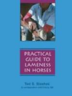
[Illustration: Fig. 41—Muscles of right leg; front view. The greater part of the long extensor has been removed. 1, 2, 3. Stumps of patellar ligaments. 4. Tuberosity of tibia. (From Sisson’s “Anatomy of the Domestic Animals.")]
The stifle joint is analagous to the knee joint of man and is to be considered an atypical ginglymus (hinge) articulation formed by the femur, tibia and patella. The ligaments are femerotibial, femeropatellar and capsular.
In addition to the usual provision for articulation of bones there are situated cartilaginous menisci between the condyles of the femur and the head of the tibia. These discs surround the tibial spine and are otherwise shaped to fit perfectly between the articular portions of the femur and tibia.
Collateral ligaments (internal and external lateral) pass from the distal end of the femur to the proximal portion of the tibia. The mesial (internal) arises from the internal condyle of the femur and is attached to a rough area below the margin of the medial (internal) condyle of the tibia. The lateral (external), shorter and thicker, arises from the depression on the lateral epicondyle and inserts to the head of the fibula.
The crucial or interosseus, anterior and posterior, are situated between the femur and tibia, and according to Smith,[34] the crucial ligaments are necessary to properly join the two bones, because of the character of the structure of the articular ends of the femur and tibia.
The femeropatella ligaments are two thin bands which reinforce the capsular ligament. They arise from the lateral aspects of the femur, just above the condyles and are inserted to the corresponding surfaces of the patella.
The patellar ligaments are three strong bands which arise from the antero-inferior surface of the patella, and are inserted to the anterior aspect of the tuberosity of the tibia.


