Scientific American Supplement, No. 460, October 25, 1884 eBook

This eBook from the Gutenberg Project consists of approximately 135 pages of information about Scientific American Supplement, No. 460, October 25, 1884.

Scientific American Supplement, No. 460, October 25, 1884 eBook

This eBook from the Gutenberg Project consists of approximately 135 pages of information about Scientific American Supplement, No. 460, October 25, 1884.
a length of several inches when fully developed, and are of an average diameter of 1/250 (0.004) inch.  They branch but sparingly, or not at all, and are characterized by consisting of a single long tube or cell, not divided by septa, as in the case of the great majority of the filamentous algae.  These tubular filaments are composed of a nearly transparent cellulose wall, including an inner layer thickly studded with bright green granules of chlorophyl.  This inner layer is ordinarily not noticeable, but it retracts from the outer envelope when subjected to the action of certain reagents, or when immersed in a fluid differing in density from water, and it then becomes distinctly visible, as may be seen in the engraving (Fig. 1).  The plant grows rapidly and is endowed with much vitality, for it resists changes of temperature to a remarkable degree. Vaucheria affords a choice hunting ground to the microscopist, for its tangled masses are the home of numberless infusoria, rotifers, and the minuter crustacea, while the filaments more advanced in age are usually thickly incrusted with diatoms.  Here, too, is a favorite haunt of the beautiful zoophytes, Hydra vividis and H. vulgaris, whose delicate tentacles may be seen gracefully waving in nearly every gathering.

REPRODUCTION IN VAUCHERIA.

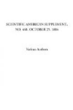
After the plant has attained a certain stage in its growth, if it be attentively watched, a marked change will be observed near the ends of the filaments.  The chlorophyl appears to assume a darker hue, and the granules become more densely crowded.  This appearance increases until the extremity of the tube appears almost swollen.  Soon the densely congregated granules at the extreme end will be seen to separate from the endochrome of the filament, a clear space sometimes, but not always, marking the point of division.  Here a septum or membrane appears, thus forming a cell whose length is about three or four times its width, and whose walls completely inclose the dark green mass of crowded granules (Fig. 1, b).  These contents are now gradually forming themselves into the spore or “gonidium,” as Carpenter calls it, in distinction from the true sexual spores, which he terms “oospores.”  At the extreme end of the filament (which is obtusely conical in shape) the chlorophyl grains retract from the old cellulose wall, leaving a very evident clear space.  In a less noticeable degree, this is also the case in the other parts of the circumference of the cell, and, apparently, the granular contents have secreted a separate envelope entirely distinct from the parent filament.  The grand climax is now rapidly approaching.  The contents of the cell near its base are now so densely clustered as to appear nearly black (Fig. 1, c), while the upper half is of a much lighter hue and the separate granules are there easily distinguished, and, if very closely watched, show an almost imperceptible motion. 

Copyrights
Project Gutenberg
Scientific American Supplement, No. 460, October 25, 1884 from Project Gutenberg. Public domain.